Embryo development days 1–6: a laboratory map
Follow a fertilised egg through pronuclei, cleavage, morula and blastocyst stages, while understanding the limits of each daily update.
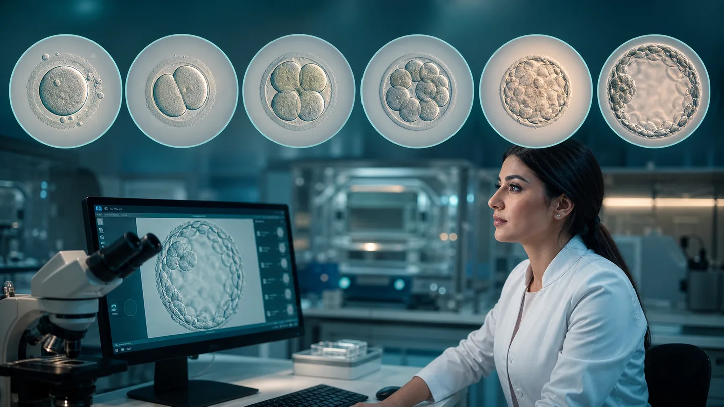
Embryo development days 1–6 describe a continuous biological process through selected laboratory observation points. Day one generally refers to the fertilisation check, followed by cell division, compaction and blastocyst formation. The day number is normally counted from egg retrieval and insemination rather than from the calendar date of an update.
The sequence is a guide, not a race with a rigid deadline. Embryos in the same culture can progress at different speeds, and slower development is not always arrest. A laboratory makes decisions according to its incubator, observation times and validated freezing criteria; direct comparison with an image online can mislead.
Days one to three: fertilisation and cleavage
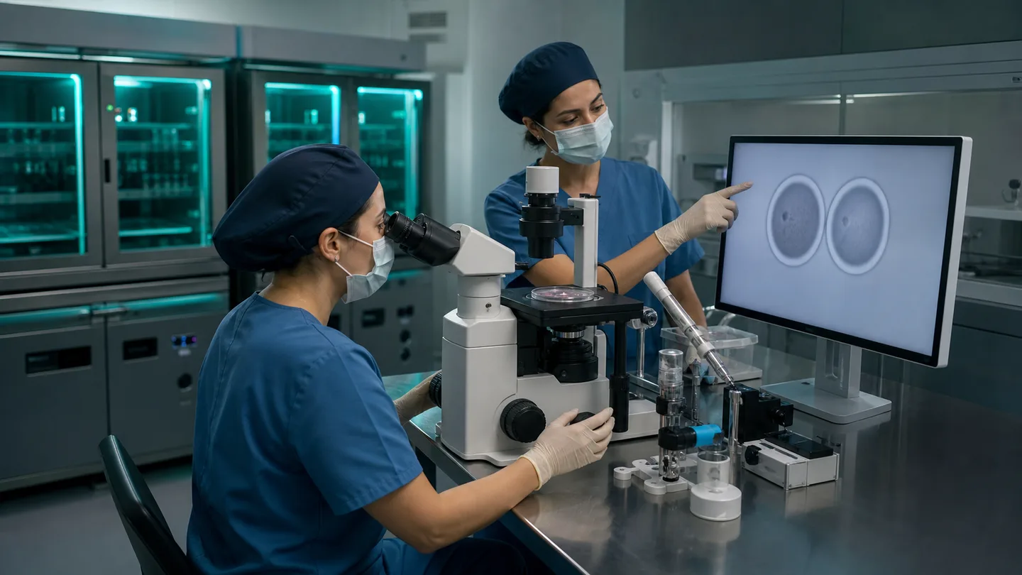
On day one, normal fertilisation is usually recorded by two pronuclei. These later disappear as the cell approaches its first mitotic division. By day two an embryo commonly contains several cells, and by day three more blastomeres may be present. Exact counts vary with the observation hour and individual pace.
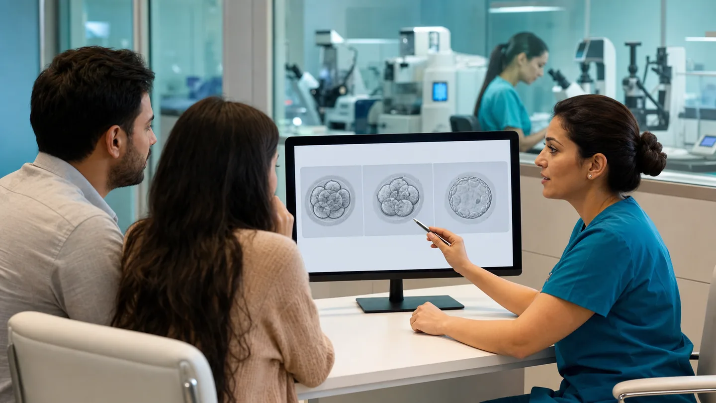
The embryologist may record cell number, relative size, fragmentation and features such as multinucleation. These are morphological observations that support relative selection. A single image does not reveal all chromosomes or the health of a future child, and an early grade cannot guarantee the final outcome.
- Day one: pronuclear check
- Day two: early cleavage
- Day three: blastomere count and appearance
Day four: morula and compaction
Around day four, boundaries between individual cells begin to become less distinct as they bind closely together. This is compaction and the embryo is described as a morula. Counting separate cells is now difficult, so assessment focuses on how complete compaction appears and any visible irregularity.
Some laboratories do not provide a routine day-four update because keeping the culture undisturbed may be preferable. A day without a telephone call does not mean development is no longer monitored. Knowing the planned communication days before retrieval can reduce uncertainty.
- Cell boundaries become compact
- Transition to the morula stage
- Fewer observations may protect incubator stability
Days five and six: blastocyst decisions
Once a fluid-filled cavity forms, the embryo is called a blastocyst. The laboratory may describe expansion, the inner cell mass and the appearance of the trophectoderm. An embryo that is not a blastocyst on day five may reach a suitable stage on day six; each laboratory applies validated time limits.
A suitable blastocyst may be transferred, vitrified or biopsied when PGT is planned. Not every fertilised cell reaches this stage, reflecting natural biological attrition. The final report should record development day, grade and outcome for each embryo, without presenting grade as a precise prediction of live birth.
- Blastocyst cavity and expansion
- Inner cell mass and trophectoderm description
- Transfer, freezing, biopsy or developmental arrest
Frequently asked questions
Is a day-six blastocyst worse than a day-five blastocyst?
Later development can be associated with some outcome differences, but the day alone does not decide an individual embryo’s fate. Grade, age, test data and clinical context also matter.
Why do I not receive an embryo image every day?
Some systems image continuously, while others require dishes to leave the incubator. The laboratory chooses observations that preserve a stable environment and provide useful information.
Ask an Övlad Payı embryologist to explain embryo development days 1–6 as a sequence from fertilisation to cleavage, morula and blastocyst. A stage map places individual numbers in a clearer context.
Trusted sources
This article is for patient education and does not replace individual medical advice.