How fertilisation is assessed: what embryologists look for
Understand the difference between collected eggs, mature eggs and normal fertilisation, including the pronuclear check and update timing.

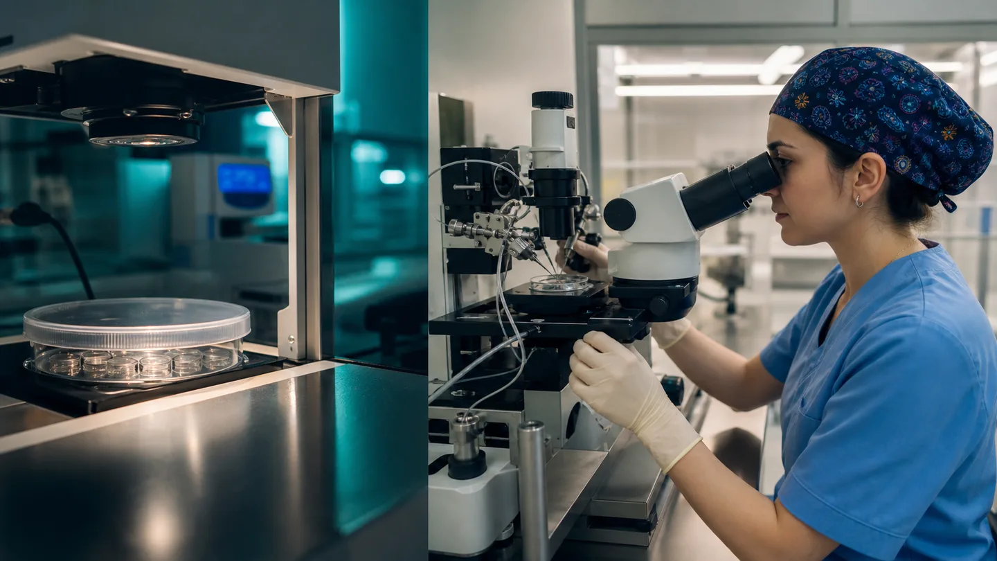
How fertilisation is assessed is the first major laboratory question after eggs and prepared sperm have been brought together. An embryologist does more than mark “fertilised” or “not fertilised”. They record egg maturity, the method used and structures visible inside the cell at the validated observation time. This becomes the starting point for following embryo development.
The number of eggs collected and the number normally fertilised will not be identical. Some cells may be immature, some may not show normal signs after exposure to sperm and a small number may show an unusual fertilisation pattern. This reduction does not automatically indicate a laboratory error and should be explained relative to the personal starting count.
Why mature eggs are counted separately
After retrieval, the cells surrounding an egg and its maturation stage are assessed. ICSI normally requires a mature metaphase-II egg; with conventional IVF, maturity is not always visualised in the same way, but the laboratory follows suitable cells. Finding immature eggs does not mean the ultrasound follicle count was necessarily wrong.
A report should separate the total collected, mature and inseminated or injected counts. That distinction may inform a later discussion about trigger timing and ovarian response, although one cycle alone cannot determine a future outcome. Ask for a plain-language explanation when abbreviations such as MII are used.
- Total number of eggs collected
- Number mature and suitable for treatment
- Number exposed to IVF or injected by ICSI
What two pronuclei mean
At the appropriate time after insemination, a normally fertilised egg usually contains two pronuclei: one associated with the egg and one with the sperm. The laboratory records this as the principal morphological sign of normal fertilisation. Pronuclei are visible during a limited window, so observation follows a validated timetable.
Zero, one, three or more pronuclei may be classified as an abnormal observation. Management follows laboratory policy, professional standards and local rules. Patients may see the abbreviation “2PN” in a report; it confirms two pronuclei were observed, but does not promise later development or implantation.
- 2PN records normal fertilisation
- Observation timing matters for interpretation
- Normal fertilisation does not guarantee continued development
When results arrive and what follows
The fertilisation check commonly takes place the morning after egg retrieval, although the communication time differs between clinics. The update should distinguish total eggs, mature eggs and normally fertilised eggs. Writing down each number and asking when the next laboratory call is expected can reduce confusion.
Normally fertilised embryos remain in controlled incubators. Later assessments follow cell division and progression towards a blastocyst. A high count on day one cannot guarantee live birth, just as a low count does not remove the potential of an individual embryo. Any forecast must include the wider clinical context.
- Record every transition in the count
- Know the timing of the next laboratory update
- Do not treat the first numbers as a guarantee
Frequently asked questions
Should every mature egg fertilise normally?
No. A proportion of mature cells may not show normal pronuclei. The rate varies with age, egg and sperm characteristics, the method and normal biological variation.
Does 2PN prove the embryo is healthy?
No. Two pronuclei indicate normal fertilisation, but cannot guarantee continued development, chromosome status, implantation or live birth.
Ask the Övlad Payı laboratory to explain how fertilisation is assessed using the total, mature and 2PN counts in sequence. A clear starting report makes later updates easier to follow calmly.
Trusted sources
This article is for patient education and does not replace individual medical advice.