Time-lapse embryo monitoring: what the camera shows
Understand how images are taken inside an incubator, the value of stable culture and why a video or algorithm cannot guarantee an outcome.
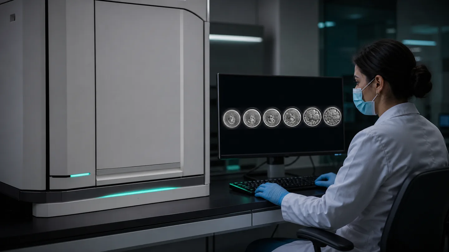
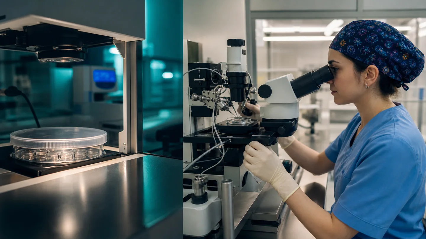
Time-lapse embryo monitoring uses a camera inside an incubator to photograph embryos at regular intervals. The images create a sequence showing the timing of cell divisions. An embryologist can review selected developmental events without removing the dish for every observation, potentially keeping culture conditions more stable.
The technology produces compelling videos, but it is not a treatment by itself. Collecting more information does not necessarily increase live birth for every patient. Some systems use algorithms to rank embryos, yet the population and laboratory in which a model was developed affect how relevant its score is elsewhere.
How the system works
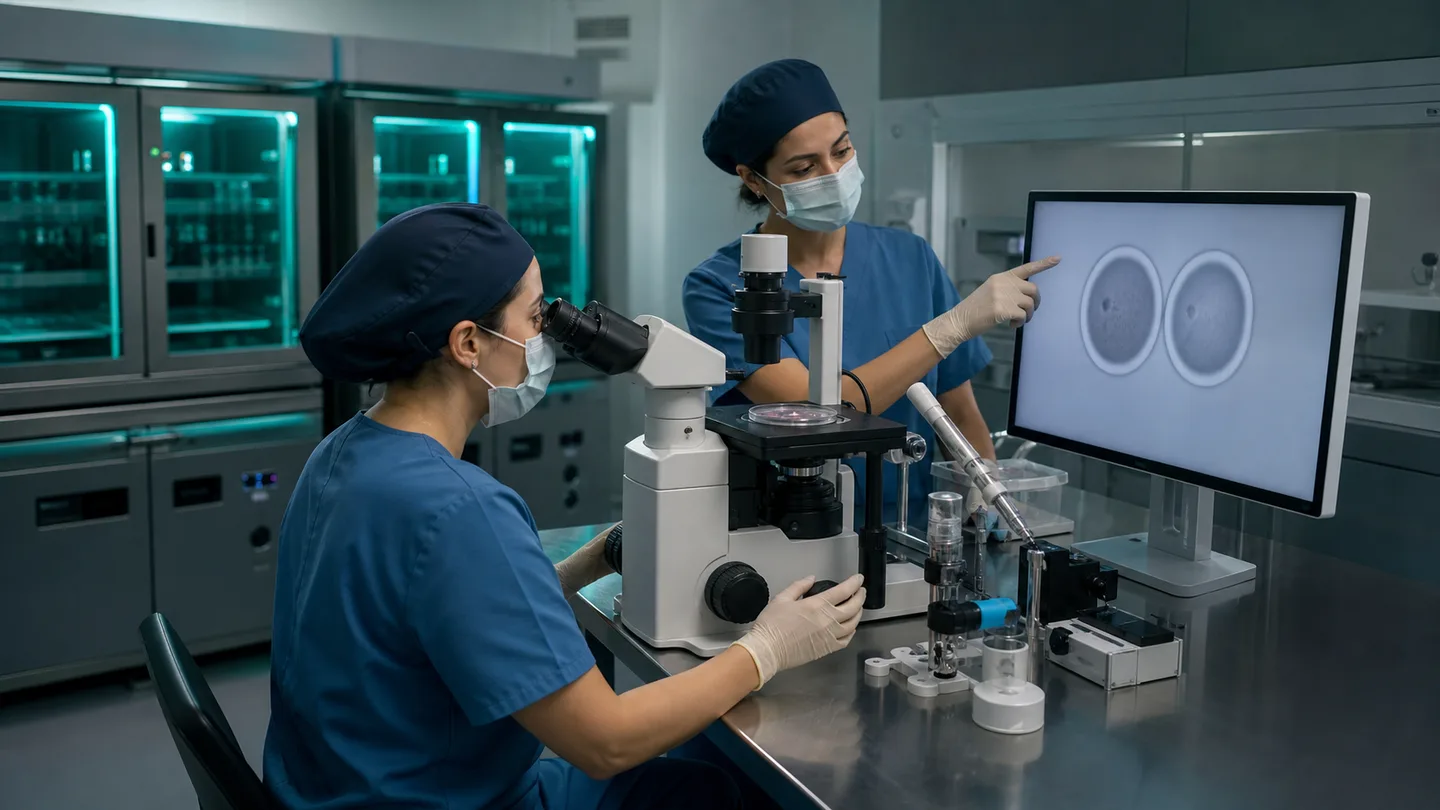
Embryos are placed in a special dish with individual wells and remain in a fixed position within the incubator. A camera can take images at several focal planes every few minutes. Software creates a sequence, while an embryologist marks events such as disappearance of pronuclei, division times and blastocyst formation.
A conventional incubator uses planned snapshot observations. Time-lapse may capture events between them, but image quality, annotation rules and culture conditions vary between systems. The camera cannot directly see an embryo’s chromosome complement or the health of a future child.
- Periodic images inside the incubator
- A timeline of cell-division events
- Fewer dish removals and potentially stable conditions
Potential value and the evidence boundary
Stable incubation can offer a practical laboratory-workflow benefit, and additional timing information may support relative embryo selection. Videos can also help communication. The clinically important question, however, is whether the technology improves live birth compared with good standard culture.
When professional assessment does not show a clear benefit for every group, the system should be presented cautiously as an add-on. An incubator with a camera cannot replace overall laboratory quality. Skilled staff, temperature and gas control, witnessing and quality systems remain fundamental.
- Additional morphokinetic information
- Uncertain live-birth advantage across all groups
- No replacement for comprehensive laboratory quality
Questions about algorithms, cost and consent
Ask whether the system is used only for incubation, for human video review or with an embryo-ranking algorithm. If an algorithm is used, ask whether it has been validated with the clinic’s results and how an embryologist reviews its suggestion. An automated score should not be the sole reason for a decision.
Understand the additional charge, access to videos, data retention and availability of standard incubation. Declining should not reduce the quality of ordinary treatment. Consent should explain potential value, uncertainty and privacy without relying on promotional language.
- Ask what exact function is being offered
- Check local validation and human oversight
- Compare cost, alternatives and data-storage terms
Frequently asked questions
Can time-lapse identify the best embryo with certainty?
No. Extra developmental information may support selection, but no video or algorithm can precisely predict implantation or a healthy live birth.
Will embryos develop less well without time-lapse?
Not necessarily. Well-managed conventional incubators support safe culture. The choice should consider the laboratory system, evidence, cost and personal preference.
Ask the Övlad Payı laboratory to explain exactly what its time-lapse embryo monitoring system does, whether an algorithm is used and what evidence supports the additional cost. Consider it a possible laboratory tool, not a guarantee.
Trusted sources
This article is for patient education and does not replace individual medical advice.